O
colo do útero está dividido em ectocérvix (porção vaginal) e endocérvix. A porção
vaginal é visível a olho nu e coberta por um epitélio escamoso estratificado
não queratinizado. O epitélio escamoso converge centralmente para orifício externo
onde encontra a endocérvix que é revestido por um epitélio colunar simples
secretor de muco. Esse epitélio mergulha no estroma subjacente formando criptas
(glândulas endocervicais). O ponto no qual o epitélio escamoso e o colunar se
encontram é denominado junção escamo-colunar ou zona de transformação.
A chamada zona de transformação é a porção da endocérvix mais próxima do
orifício externo do canal cervical e, portanto, limítrofe com a ectocérvix.
Nesta região é comum a ocorrência de metaplasia escamosa.
Translation: Cancer
of the cervix, also known as cervical, takes many years to develop. The cell changes that can trigger cancer are easily discovered on Pap
(also known as Pap), so it is important your periodical. The main change that
can lead to this type of cancer is infection with human papillomavirus, HPV,
with some high-risk subtypes and related malignancies.
The cervix is divided into ectocervix (vaginal
portion) and endocervix. The vaginal portion is visible to the naked eye,
covered by a non-keratinized stratified squamous epithelium. The squamous
epithelium converge centrally to external orifice where it meets the endocervix
is lined by a simple columnar epithelium secreting mucus. This plunges into the
stroma underlying the epithelium forming crypts (endocervical glands). The
point at which the squamous epithelium and the columnar meet is called the
squamo-columnar junction or transformation zone.
The call processing area is the portion closest
to the endocervix external os of the cervical canal and thus bordering the
ectocervix. This region is a common occurrence of squamous metaplasia.
 |
| Anatomia do útero - Translation: Anatomy
of the uterus |
 |
| Modelo esquemático do desenvolvimento da neoplasia intra-epitelial cervical (NIC) - Translation: Schematic model of development of
cervical intraepithelial neoplasia (CIN) |
4x
Visualização detalhada(Details)
Na imagem,nota-se a presença do ectocervice(região do colo do
útero que fica voltada para o cana vaginal),caracterizado por
apresentar um epitélio pavimentoso estratificado(seta amarela).Também se nota
uma presença marcante de angiogênese(setas azuis).
Translation: In the picture, note the presence
of the ectocervix (cervical region that faces the vaginal canal), characterized
by presenting stratified squamous epithelium (yellow arrow). Also note a
marked presence from angiogenesis (blue arrows).
10x
Epitélio normal do ectocérvice(seta azul),e
presença de acantose – espessamento do epitélio(seta preta).
Translation: Normal epithelium of the ectocervix (blue arrow), and presence of acanthosis - thickening of the epithelium (black arrow).
Translation: Normal epithelium of the ectocervix (blue arrow), and presence of acanthosis - thickening of the epithelium (black arrow).
40x
Visualização detalhada(Details)
Na imagem,nota-se presença em
alguma áreas do ectocérvice espongiose(setas pretas),degeneração hidrópica(setas amarelas) e exocitose(setas azuis).
Translation: In the picture, note the presence in some areas of the ectocervix, spongiosis (black arrows), hydropic degeneration (yellow arrows) and exocytosis (blue arrows).
Translation: In the picture, note the presence in some areas of the ectocervix, spongiosis (black arrows), hydropic degeneration (yellow arrows) and exocytosis (blue arrows).

4x
Visualização detalhada(Details)
Endocérvice – apresenta duas características marcantes :
1-o epitelio de revestimento:epitélio cilíndrico simples mucoso
2 – glândulas exócrinas tubulares(glândulas endocervicais) compostas de epitélio cilíndrico simples mucoso(ambos
representados pelas setas amarelas)
Translation: Endocervix - has two remarkable
features:
1-the epithelium lining: simple cylindrical
epithelium mucous
2 - tubular exocrine glands (glands
endocervical) composed of simple cylindrical epithelium mucous (both
represented by yellow arrows).
40x
Visualização detalhada(Details)
Na imagem,nota-se a glândula endocervical,com seu epitélio cilíndrico simples
mucoso(seta amarelas).
Translation: In the picture, it is noticed the endocervical gland, with its simple cylindrical epithelium mucus (yellow arrow).
Translation: In the picture, it is noticed the endocervical gland, with its simple cylindrical epithelium mucus (yellow arrow).
40x
Visualização detalhada(Details)
Na imagem,nota-se glândula
endocervical com um epitélio normal(seta azul) que sofre uma metaplasia,se tornando
estratificado – metaplasia da glândula endocervical(seta amarela).Também nota-se a
presença de exocitose.
Translation: In the image, there is endocervical gland with a normal epithelium (blue arrow) that suffers a metaplasia, becoming stratified - metaplasia of endocervical gland (yellow arrow). Also note the presence of exocytosis.
Translation: In the image, there is endocervical gland with a normal epithelium (blue arrow) that suffers a metaplasia, becoming stratified - metaplasia of endocervical gland (yellow arrow). Also note the presence of exocytosis.
40x
Visualização detalhada(Details)
Visualização detalhada(Details)
Na imagem,nota-se a presença de uma transformação do epitélio endocervical para epitélio
metaplásico(seta preta) –
notar também presença de displasia no epitélio transformado,com presença de pleomorfismo celular e
nuclear,hipercromatismo nuclear em toda a camada do epitélio metaplasico(círculo
azul).Nota-se exocitose(seta amarela).
Translation: In the picture, note the presence of a transformation of the endocervical epithelium to metaplastic epithelium (black arrow) - also note the presence of dysplasia in the transformed epithelium with the presence of nuclear and cellular pleomorphism nuclear, hyperchromatic in all layer of the metaplastic epithelium (circle blue). Note to exocytosis (yellow arrow).
Translation: In the picture, note the presence of a transformation of the endocervical epithelium to metaplastic epithelium (black arrow) - also note the presence of dysplasia in the transformed epithelium with the presence of nuclear and cellular pleomorphism nuclear, hyperchromatic in all layer of the metaplastic epithelium (circle blue). Note to exocytosis (yellow arrow).
Na imagem,nota-se a presença de um outro achado histopatologico -> metaplasia escamosa do endocervice.A presença das
glândulas endocervicais(cabeças de seta),nos garante que esta área se
trata do endocervice,o qual
deveria apresentar epitélio
cilíndrico simples mucoso.No entanto,devido ao fato de ter sofrido uma metaplasia escamosa,seu epitelio se tornou pavimento
estratificado com presença de querato-hialina(seta azul).
Translation: In the picture, note the presence of another histopathological findings -> endocervice.A presence of squamous metaplasia of the endocervical glands (arrowheads), assures us that this area it is the endocervix, which should present simple cylindrical epithelium mucous. However, due to having suffered a squamous metaplasia, its epithelium became stratified pavement with the presence of keratohyalin (blue arrow)
Translation: In the picture, note the presence of another histopathological findings -> endocervice.A presence of squamous metaplasia of the endocervical glands (arrowheads), assures us that this area it is the endocervix, which should present simple cylindrical epithelium mucous. However, due to having suffered a squamous metaplasia, its epithelium became stratified pavement with the presence of keratohyalin (blue arrow)
40x
Visualização detalhada(Details)
Visualização detalhada(Details)
Em maior detalhe,presença de querato-hialina(seta azul).
Translation: In more detail the presence of keratohyalin (blue arrow).
Translation: In more detail the presence of keratohyalin (blue arrow).
10x
Visualização detalhada(Details)
Visualização detalhada(Details)
Na imagem,nota-se a presença do epitelio metaplasico com querato-hialina(que também apresenta displasia severa,pois houve alterações em
todas as camadas do epitélio)[seta vermelha].Nota-se também a invasão de um
grupo de céluas neoplasicas,proveniente do epitélio metaplasico,que romperam a membrana basal e estão invadindo o
conjuntivo adjacente(setas amarelas).Presença de ilhas de células neoplásica
que já se destacaram do epitélio(seta azuis).A área em questão é o endocervice,devido
presença das glâdulas endocervicais(cabeça de setas).
Translation: In the picture, note the presence of metaplastic epithelium with keratohyalin (also shows severe dysplasia because there have been changes in all layers of the epithelium) [red arrow]. Note also the invasion of a group of neoplastic cells, coming from metaplastic epithelium, which pierced the basal membrane and are invading the adjacent conjunctive (yellow arrows). Presence of islands of neoplastic cells already detached from the epithelium (blue arrow). The area in question is the endocervix, because the presence of endocervical glands (arrow head).
Translation: In the picture, note the presence of metaplastic epithelium with keratohyalin (also shows severe dysplasia because there have been changes in all layers of the epithelium) [red arrow]. Note also the invasion of a group of neoplastic cells, coming from metaplastic epithelium, which pierced the basal membrane and are invading the adjacent conjunctive (yellow arrows). Presence of islands of neoplastic cells already detached from the epithelium (blue arrow). The area in question is the endocervix, because the presence of endocervical glands (arrow head).
Grande ilha de células neoplasicas.
Translation: Great Island of neoplastic cells.
Translation: Great Island of neoplastic cells.
Na imagem,nota-se presença de um epitélio completamente metaplasico(com displasia
severa)
no endocervice,repleto de pleomorfismo celulares e
nucleares(setas vermelhas),hipercromatismo nuclear(setas
azuis),além de figuras de mitose bizarras(seta amarela).No quadrado amarelo há
mais das mesmas alterações do quadrado azul.
Translation: In the picture, note the presence of a completely metaplastic epithelium (severe dysplasia)in the endocervix, filled with cellular pleomorphism and nuclear (red arrows), hyperchromatic nuclear (blue arrows), and bizarre mitotic figures (yellow arrow). In the yellow box for more of the same changes the blue square.
Translation: In the picture, note the presence of a completely metaplastic epithelium (severe dysplasia)in the endocervix, filled with cellular pleomorphism and nuclear (red arrows), hyperchromatic nuclear (blue arrows), and bizarre mitotic figures (yellow arrow). In the yellow box for more of the same changes the blue square.
Epitélio metaplásico(com displasia
severa) notar o hipercromatismo e pleomorfismo celular e nuclear(setas amarelas).Um achado interessante
nessa imagem é a presença de corpos apoptóticos(seta azul) – não é pelo fato de um tumor apresentar células
imortais,que significa que nenhuma possa morrer.Pelo contrário,lembre-se que as
alterações que as células sofrem além de lhes concederem vantagens,também não
podem ser letais,caso contrário,ela entra em apoptose,como foi,possivelmente o
caso com esta célula apoptótica.
Translation: Metaplastic epithelium (severe dysplasia) notice hyperchromatism and cellular pleomorphism and nuclear (yellow arrows). An interesting finding in this image is the presence of apoptotic bodies (blue arrow) - it is not because of a tumor presenting cells immortal, which means no can die. Instead, remember the changes that cells undergo as well as granting them advantages, can not be lethal, otherwise it goes into apoptosis, as was possibly the case with this cell apoptosis.
Translation: Metaplastic epithelium (severe dysplasia) notice hyperchromatism and cellular pleomorphism and nuclear (yellow arrows). An interesting finding in this image is the presence of apoptotic bodies (blue arrow) - it is not because of a tumor presenting cells immortal, which means no can die. Instead, remember the changes that cells undergo as well as granting them advantages, can not be lethal, otherwise it goes into apoptosis, as was possibly the case with this cell apoptosis.
Na imagem,nota-se a presença de
um achado marcante,o cisto de Naboth(seta amarela).Este cisto,nada mais é,do que uma glândulas
endocervical que foi impedida de expelir seu produto,pelo fato do epitélio
endocervical ter sofrido uma metaplasia escamosa(seta azul),e com isso ocorreu o acúmulo de muco em
seu interior,dando esta forma abalonada e gigante.
Translation: In the picture, note the presence of a remarkable finding, Naboth cyst (yellow arrow). Cyst This is nothing more than a endocervical glands that was prevented from expelling their product, because the endocervical epithelium suffered a squamous metaplasia (blue arrow), and this occurred with the accumulation of mucus inside, leading him to this giant form.
Translation: In the picture, note the presence of a remarkable finding, Naboth cyst (yellow arrow). Cyst This is nothing more than a endocervical glands that was prevented from expelling their product, because the endocervical epithelium suffered a squamous metaplasia (blue arrow), and this occurred with the accumulation of mucus inside, leading him to this giant form.
Na imagem,com detalhes observa-se a compressão do muco sintetizado pela glândula endocervical(agora cisto
de Naboth).Notem que o epitélio que antes era
cilíndrico,ficou cúbico(seta azul)e em algumas regiões da glândula
pavimentoso(seta preta).
Translation: Pictured in detail, there is the compression exerted by mucus synthesized by endocervical gland (now Naboth cyst). Note that the cylindrical epithelium that was before it became cubic (blue arrow), and in some regions of the gland, squamous (black arrow).
Translation: Pictured in detail, there is the compression exerted by mucus synthesized by endocervical gland (now Naboth cyst). Note that the cylindrical epithelium that was before it became cubic (blue arrow), and in some regions of the gland, squamous (black arrow).
Angiogênese no endocérvice.
Translation: Angiogenesis in the endocervix
Translation: Angiogenesis in the endocervix
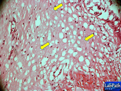
Presença de exsudato(setas amarelas).
Translation: Exudate (yellow arrows)
Translation: Exudate (yellow arrows)
Infiltrado inflamatório linfoplasmomcitário no endocérvice.
Translation: Lymphocytic inflammatory infiltrate in the endocervix
Translation: Lymphocytic inflammatory infiltrate in the endocervix
Para visualizar os vídeos sobre câncer cervical click nos links abaixo
Translation: To view the videos on cervical cancer click on the links below:
Translation: To view the videos on cervical cancer click on the links below:
Módulo 2: Replicação do Papiloma Vírus Humano e disfunção do ciclo celular(Module 2: Human
Papilloma Virus replication and cell cycle dysfunction)
Módulo 3: Neoplasia Intra-epitelial Cervical(Cervical Intraepithelial Neoplasia)
Módulo 4: Carcinoma Invasivo(Invasive Carcinoma)
Fonte:
Robbins & Cotran, Bases Patológicas das Doenças, 7ª ed.


Nenhum comentário:
Postar um comentário