segunda-feira, 14 de maio de 2012

Fibroma/Fibroma

Fibroma é um tumor do tipo benigno originado a partir de células fibroblásticas, sendo composto unicamente de elementos fibrosos.

Translation: Fibroma is a benign type tumor originated from fibroblastic cells, being composed only of fibrous elements.


 40x
Na imagem,nota-se uma grande quantidade de fibras de colágeno produzidas,além da presença de inúmeros fibroblastos morfologicamente próximos da normalidade.Notar também que as fibras nesse tumor se encontram organizadas em uma mesmas direção(círculos azuis).

Translation: In the picture, there is a large amount of collagen fibers produced and the presence of numerous fibroblasts morphologically close to normal. Note also that fibers in this tumor are arranged in a same direction (blue circles)

 40 x
Na imagem,há a presença de angiogênese(seta azuis) e um leve infiltrado inflamatório(círculo azul).
Translation: In the image, there is the presence of angiogenesis (blue arrow) and a mild inflammatory infiltrate (blue circle). 

 40x
Na imagem,nota-se presença de angiogênese(setas azuis) e formação de edema – afastamento das estruturas do parênquima(círculo azul).Uma informação importante que prova que a estrutura recém comentada se trata de um edema,é pelo fato de que há uma grande quantidade de vênulas(setas azuis),as quais são tidas na histologia como os vaso mais permeáveis(em comparação aos outros).Além disso,a hipótese de ser uma necrose é descartada,pois uma de suas características principais é a eosinofilia que ela provoca devido a presença de proteínas desnaturadas.

Translation: In the image, there is angiogenesis (blue arrows) and edema formation - remoteness of structures parenchyma (blue circle). Important information that proves that the structure recently commented this is an edema, is the fact that there are a lot of venules (blue arrows), which are taken as the vessel histology in more permeable (compared to others). Furthermore, the hypothesis is discarded necrosis be one, since one of its main features is that it causes eosinophilia due to the presence of denatured proteins.

 40x
Na imagem,é possivel ver a presença de desmoplasia – fibras mais coradas intensamente(setas azuis) e fibroblastos morfologicamente próximos da normalidade.
Translation: In the image, it is possible to see the presence of desmoplasia - fibers more intensely stained (blue arrows) and fibroblasts morphologically close to normal.


Carcinoma de Esôfago/Carcinoma of the esophagus

       O carcinoma de células escamosas é uma forma de câncer histologicamente distinta. Ele surge da multiplicação descontrolada de células do epitélio ou de células que apresentam características citológicas ou arquitetura tecidual particulares de diferenciação de células escamosas, tais como a presença de queratina, tramas de tonofilamentos ou desmossomos.
   O carcinoma de células escamosas ocorre, como uma forma de câncer, em diversos tecidos, incluindo os lábios, a boca, o esôfago, a bexiga urinária, a próstata, os pulmões, a vagina, o colo do útero, dentre outros. Apesar de compartilhar o nome "carcinoma de células escamosas" em diferentes partes do corpo, as patologias podem apresentar grandes diferenças de sintomas apresentados, história natural,prognóstico e resposta ao tratamento.
        O carcinoma de células escamosas do esôfago(ou  carcinoma esofágico) apresenta correlação ao consumo do álcool e do tabaco(porém ha regiões do mundo onde o consumo destes é nulo - muçulmanos por exemplo - e ainda assim sofrem pelo carcinoma esofágico).Deficiências nutricionais,hidrocarbonetos policíclicos,nitrosaminas e outro compostos mutagênicos,tais como aqueles encontrados em alimentos contaminados por fungos,devem ser considerados.(A patogenia ainda é indefinida,porém a perda de vários genes supressores de tumor,incluindo p53 e p16/INK4a,está envolvida.
             O Carcinoma esofágico se inicia como uma lesão in situ chamada displasia escamosa(Neoplasia intraepitelial ou carcinoma in situ).Ao longo do anos pode se  tornar uma massa tumoral poliploide ou exofítica que se projetam para dentro da luz do lúmen causando sua obstrução.Os outro tumores ou são lesões ulceradas ou difusamente infiltrativas que se espalham dentro da parede esofágica e causa espessamento,enrijecimento e estreitamento luminal.Esta podem invadir estruturas circundantes,incluindo a árvore brônquica,causando pneumonia;a aorta causando exsanguinação catastrófica;ou mediastino ou pericárdio. - Fonte : Robbins & Cotran - Patologia : Bases patológicas das doenças - 8ª ed.

Translation: The squamous cell carcinoma is a form of cancer histologically distinct. It arises from the uncontrolled proliferation of epithelial cells or cells which have cytological features or particular tissue architecture squamous cell differentiation, such as the presence of keratin tonofilaments frames or desmosomes.
   The squamous cell carcinoma occurs as a form of cancer in various tissues, including the lips, mouth, esophagus, urinary bladder, prostate, lungs, vagina, cervix, among others. Despite sharing the name "squamous cell carcinoma" in different parts of the body pathologies may exhibit large differences in symptoms, natural history, prognosis, and response to treatment.
        The squamous cell carcinoma of the esophagus (esophageal carcinoma or) correlates the consumption of alcohol and tobacco (though ha regions of the world where the consumption of these is null - eg Muslims - and still suffer by esophageal carcinoma). Nutritional deficiencies, polycyclic hydrocarbons, nitrosamines and other mutagenic compounds, such as those found in food contaminated by mold, must be considered. (The pathogenesis is still unclear, but the loss of several tumor suppressor genes including p53 and p16/INK4a is involved.
             The esophageal carcinoma starts as an injury call-situ squamous dysplasia (intraepithelial neoplasia or carcinoma in situ). Throughout the years can become a polyploid or exophytic tumor mass protruding into the lumen of light causing your other obstrução.Os or tumors are diffusely infiltrative or ulcerated lesions that spread within the esophageal wall and causes thickening, hardening and narrowing luminal.Esta may invade surrounding structures, including the bronchial tree, causing pneumonia, the aorta causing catastrophic exsanguination, or mediastinum or pericardium.             
           
 4x
Na imagem,pode-se notar na região do epitélio da mucosa esofágica uma displasia severa(seta negra),a qual é caracterizada por comprometer todas as camadas do epitélio(também pode ser chamada de carcinoma in situ,pelo fato de se encontrar confinado,ainda,no epitélio, não havendo ruptura da membrana basal);As setas amarelas apontam para as cristas do epitélio,mostrando que a membrana basal continua intacta e as células epiteliais continuam confinadas ao epitélio.Reparar na hialinização do epitélio(seta azul)provocada pela presença de degeneração hidrópica e espongiose;No círculo nota-se a presença de infiltrado inflamatório do tipo linfoplasmocitário(crônico). 
Translation: In the image, it can be noted in the region of the esophageal mucosal epithelium severe dysplasia (black arrow), which is characterized by compromising all layers of the epithelium (also can be called carcinoma in situ, because finding still confined in the epithelium, and no rupture of the basal membrane); yellow arrows indicate the crests of the epithelium, showing that remains intact basal membrane and epithelial cells remain confined to the epithelium. Repair hyalinization in the epithelium (blue arrow) caused by the presence of hydropic degeneration and spongiosis; circle No note is the presence of inflammatory infiltrates lymphoplasmocytarian (chronic).

 40x
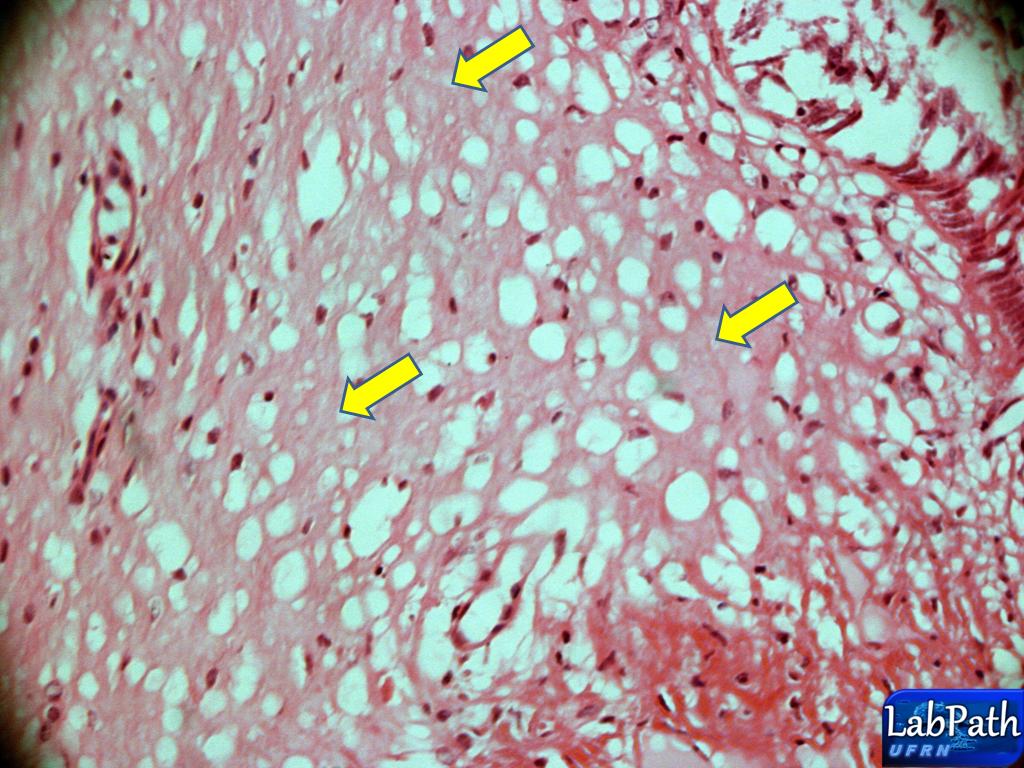
Na imagem é possível ver degeneração hidrópica – acúmulo de água intracelular(setas azuis),espongiose – acúmulo de água intercelular(setas vermelhas),presença de exocitose – células inflamatórias que invadem o epitélio(seta amarela) e hipercromatismo nuclear(setas pretas).
Translation: In the picture is possible to see hydropic degeneration - accumulation of intracellular water (blue arrows), spongiosis - accumulation of intercellular water (red arrows), presence of exocytosis - inflammatory cells that invade the epithelium (yellow arrow) and hyperchromatic nuclear (black arrows).

 4x
Geralmente,ao lado de um carcinoma in situ,nota-se o epitélio neoplásico invadindo o tecido adjacente(seta preta)-nessa região de invasão,a membrana basal já foi rompida pela produção de metaloproteases provenientes das  células invasoras.Presença de ulcerção(seta cinza) – a ulceração é um dos meios pelos quais o carcinoma  esofágico  se utiliza para invadir os tecidos adjacentes;a difusão das células neoplásicas pelo epitélio para chegar ao tecido adjacente também é um outro meio utilizado por este adenocarcinoma.Presença de angiogênese – desencadeada pela produção de fatores de crescimento que estimulam o desenvolvimento de neovasos(por exemplo o VEGF)para que haja a vascularização do tumor e este tenha o aporte sanguíneo necessário para seu desenvolvimento – tanto células neoplásicas quanto células normais (macrófagos e fibroblastos por exemplo) produzem essa moléculas(cabeças de setas),presença de infiltrado inflamatório crônico(seta amarela).
Translation: Usually, beside a carcinoma in situ, it is noted invading neoplastic epithelium adjacent tissue (black arrow) in the region of invasion, the basement membrane has been ruptured by production of metalloproteases derived from invading cells. Presence of ulcerations (gray arrow) - the ulceration is one means by which uses esophageal carcinoma to invade surrounding tissue, the spread of the neoplastic epithelial cells to reach the surrounding tissue is also another medium used for this adenocarcinoma.Presença angiogenesis - triggered by the production of growth factors that stimulate the development of neovascularization (e.g., VEGF) so that there is tumor vascularization and this has the blood supply necessary for their development - both neoplastic cells as normal cells (fibroblasts and macrophages for example) produce such molecules (arrowheads), presence of chronic inflammatory infiltrate (yellow arrow).

 40x
Na imagem,pode-se notar presença de exocitose(setas azuis),hipercromatismo nuclear(setas pretas),pleomorfismo celular e nuclear(setas vermelhas) e presença se células neoplásicas saindo do epitélio de origem e invadindo o conjuntivo adjacente(seta amarelas).
Translation: In the image, one can notice the presence of exocytosis (blue arrows), hyperchromatic nuclear (black arrows), cellular pleomorphism and nuclear (red arrows) and presence of neoplastic cells were exiting the epithelium of origin and invading the adjacent conjunctive (yellow arrow).

 10x
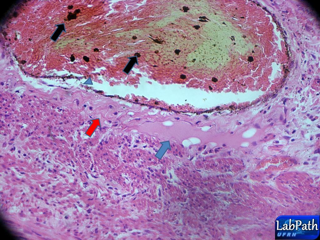
Presença de grande ulceração no epitélio(seta azul) culminado na invasão do conjuntivo adjacente por um grande grupo de células neoplásicas do epitélio – reparar na perda de membrana basal(seta preta),grande produção de fibras de colágeno(desmoplasia) – células neoplásicas estimulam células do parênquima a produzirem colágeno).
Translation: Presence of great ulceration in the epithelium (blue arrow) culminating in the invasion of adjacent conjunctive by a great group of neoplastic epithelial cells - notice the loss of basal membrane (black arrow), great production of collagen fibers (desmoplasia) - neoplastic cells stimulate parenchyma cells to produce collagen).

 10x
Na imagem,nota-se presença de ilha de células neoplásicas que invadiram o tecido conjuntivo(setas pretas) e também de disceratoses – células neoplásicas que produzem queratina individualmente(setas azuis).Ambas características citadas rementem a um bom prognóstico para o paciente,uma vez que mostram o quanto as células neoplásicas estão próximas da normalidade,ou seja,ainda apresentam um bom grau de diferenciação.
Translation: In the picture, note the presence of the island of neoplastic cells that have invaded the conjunctive tissue (black arrows) as well as dyskeratosis - neoplastic cells that produce keratin individually (blue arrows). Both mentioned characteristics are related to a good prognosis for patients, since they show how much the neoplastic cells are close to normal, as well as the degree of differentiation show good.

 10x
Na imagem,nota-se a presença de uma ilha de células neoplásica(seta amarela)sendo atacada por uma grande quantidade de células provenientes do infiltrado inflamatório linfoplasmocitário(seta vermelha).Também a presença de desmoplasia(setas azuis).
Translation: In the picture, note the presence of an island of neoplastic cells (yellow arrow) being attacked by a large amount of cells from the lymphocytic inflammatory infiltrate (red arrow). Also the presence of desmoplasia (blue arrows).
 40x
Na imagem,nota-se presença de ilha de células neoplásica(seta preta) com presença de perola córnea – células neoplásicas que passaram a produzir queratina juntas(seta azul).Também significa um bom prognostico pelo mesmo motivo citado acima.
Translation: In the picture, note the presence of neoplastic cells Island (black arrow) with the presence of corneal pearl - neoplastic cells that produce keratin together (blue arrow). Means also a good prognosis for the same reason mentioned above.

 40X
Visualização detalhada(Details)
Na imagem nota-se intensa angiogênese com formação de neovasos(seta azuis) e células neoplásicas que invadiram o conjuntivo(setas amarelas) cercadas pelo infiltrado inflamatorio(círculo).
Translation: Pictured is noted intense angiogenesis with formation of new vessels (blue arrow) and neoplastic cells that have invaded the tissue (yellow arrows) surrounded by inflammatory infiltrate (circle).

Presença de hemorragia.
Translation: Presence of hemorrhage.


sábado, 12 de maio de 2012

Apendicite aguda/Acute appendicitis

A inflamação aguda é uma rápida resposta do hospedeiro que serve para levar leucócitos e proteínas plasmáticas,tais como anticorpos,para os locais de infecção ou tecido injuriado.A inflamação aguda tem 3 principais componentes: (1)alterações no calibre vascular que levam a um aumento do fluxo sanguíneo;(2) mudanças estruturais na microvasculatura que permitam a saída das proteínas plasmáticas e os leucócitos da circulação(predominantemente polimorfos nucleares)  e (3) emigração de leucócitos da microcirculação,seu acúmulo no foco da injúria e sua ativação para eliminar o agente agressor.
O apêndice é um divertículo verdadeiro normal do ceco que está sujeito a inflamação crônica ou aguda,sendo esta mais prevalente em adolescentes e jovens  adultos.Sabe-se que a apendicite aguda se inicia pelo aumento progressivo na pressão intraluminal que compromete o fluxo venoso de saída.A injúria isquêmica e a estase do conteúdo luminal,que favorecem a proliferação bacteriana,disparam uma resposta inflamatória incluindo edema tissular,e infiltração neutrofílica.
Fonte : Robbins & Cotran - Patologia : Bases Patológicas das Doenças - 8ª ed.

Translation : Acute inflammation is a rapid host response that serves to carry leukocytes and plasma proteins, such as antibodies, to sites of infection or injured tissue. Acute inflammation has three main components: (1) changes in vascular caliber leading to an increased blood flow and (2) structural changes in the microvasculature enabling the output of plasma proteins and leukocytes from the circulation (predominantly polymorph nuclear) and (3) emigration of leukocytes in the microcirculation, their accumulation in the focus of injury and their activation to eliminate the offending agent.

The appendix is ​​a true diverticulum normal cecum which is subject to  chronic  or acute inflammation, which is more prevalent in adolescents and young adults. It is known that acute appendicitis begins with the progressive increase in intraluminal pressure, which compromises the venous flow output. Ischemic injury and stasis of luminal contents, which favors bacterial proliferation, triggers an inflammatory response including tissue edema, and neutrophil infiltration.


  4x
Notar na imagem a grande destruição provocada pela inflamação aguda(que tem um início abrupto e severo),percebendo-se um parênquima totalmente alterado(estrela azul),desorganização do nódulos linfáticos(setas pretas);notar também erosão na mucosa do apêndice(seta azul) – estrela amarela indica a luz do apêndice.
Translation: Notice in the picture the vast destruction caused by acute inflammation (which has a sudden onset and severe), realizing a completely modified parenchyma (blue star), disorganization of lymph nodes (black arrows); note also erosion in the mucosa of the appendix (arrow blue) - light yellow star indicates the lumen  of the appendix.


 10x
Nota na imagem áreas de hemorragia– reparar na refringência e na presença de hemossiderina -  na mucosa do apêndice(círculos);reparar na quantidade de plasma liberado devido ao processo inflamatório(setas azuis).O quadrado preto mostra a desorganização dos nódulos linfáticos associados à mucosa.
Translation: Note in the image extensive hemorrhages - notice the refringence and the presence of hemosiderin - in the mucosa of Appendix (circles); notice the released amount of plasma due to inflammation (blue arrows). The black square shows the disorganization of the lymph nodes associated to mucosa.


 10x
Notar na imagem as setas pretas que indicam a desorganização do MALT,as amarelas indicam tecido adiposo e as azuis indicam deposição de colágeno(fibrose).As estrela amarelas indicam liberação de plasma e fibrina.
Translation : Notice in the image that the black arrows indicate the disorganization of MALT, yellow arrows indicate adipose tissue and the blue arrows indicate collagen deposition (fibrosis). The yellow star indicates and release of plasma fibrin.


10x
Notar na imagem uma das característica mais importantes desta lâmina: o espessamento(estrela preta) da serosa(cabeça de seta azul) e a presença de inúmeros vasos congestos(estrelas azuis pequenas). A estrela azul maior mostra a camada muscular circular interna e a amarela indica a longitudinal externa.
Translation:  Notice in the image one of the most important feature of this blade: thickening (black star) serous of the layer (blue arrowhead) and the presence of numerous congested vessels (small blue stars). A larger blue star shows the internal circular muscle layer and yellow indicates the outer/extern  longitudinal muscle layer.


 40x
Notar na imagem um vaso congesto(hiperemiado) afrouxa devido a quantidade de hemácias e plasma(cabeça de seta azul) liberando fibrina(seta vermelha) e plasma(seta azul).As setas pretas indicam a presença de hemossiderina.
Translation: Notice in the image a vase congested (hyperemic) loosing because the amount of red blood cells and plasma (blue arrowhead) releasing fibrin (red arrow) and plasma (blue arrow). The black arrows indicate the presence of hemosiderin.


  4x
Notar que o infiltrado inflamatório já atingiu as camadas musculares.
Translation: Note that the inflammatory infiltration has reached the muscle layers.


 40x
Notar a presença de PMNS,(cabeça de setas);as estrela mostra exsudato plásmatico.
Translation: Note the presence of PMNs (arrow head); star shows the plasma exudate.


Metástase de melanoma


Esquema de metástase


Aumento de 4x
Na imagem,nota-se um parênquima normal do tecido hepático,com seus cordões hepático,sinusóides hepáticos e veia centro-lobular


Aumento 40x
Mais detalhes da imagem anterior,onde se notam a presença dos cordões de hepatócitos(setas azuis) e dos sinusóides hepáticos(cabeças de seta)


Aumento 40x
Na imagem,nota-se a presença de um tumor secundário,que levou a morte os cordões hepáticos(Linha delimitante azul)


Aumento 40x
Na imagem,notam-se melanócitos anaplásicos(cabeça de setas) que metastisaram  para o lóbulo hepático,provocando a apoptose do hepatócitos(círculo azul) .

Aumento de 40x
Na imagem,notar a presença de melanócitos anaplásicos(cabeça de seta) e cordões hepáticos sofrendo apoptose(círculo)

Aumento 40x
Na imagem,nota-se presença de infiltrado linfoplamocitário(círculo azul) tentando barrar a progressão da invasão por parte dos melanócitos neoplásicos.

Aumento de 40x
Na imagem,nota-se o infiltrado linfoplamocitário combatendo os melanócitos invasores


Aumento de 40x
Na imagem,nota-se uma intensa produção de fibras de coláneo – fibrose –(círculo)como tentativa de barrar a progressão da invasão tumoral – notar presença de vários fibroblastos(cabeça de setas) e alguns melanócitos que forma barrado pela produção de fibras(quadrado)

Aumento de 10x
Na imagem,nota-se melanócitos neoplasicos seguindo em direção ao parênquima normal,invadindo-o(círculos)

Aumento de 40x
Mais detalhes da invasão dos melanócitos ao parênquima normal(setas)

Aumento de 40x
Presença de áreas de hemorragia

Aumento de 40x
Presença de um exsudato fibrinoso(círculo azul)

Aumento de 10x
Devido a presença de hemorragia vamos encontrar a presença de hemossiderina(cabeça de setas)


domingo, 6 de maio de 2012

Carcinoma de colo uterino/Carcinoma of the cervix

O câncer do colo do útero, também chamado de cervical, demora muitos anos para se desenvolver. As alterações das células que podem desencadear o câncer são descobertas facilmente no exame preventivo (conhecido também como Papanicolaou), por isso é importante a sua realização periódica. A principal alteração que pode levar a esse tipo de câncer é a infecção pelo papilomavírus humano, o HPV, com alguns subtipos de alto risco e relacionados a tumores malignos.
O colo do útero está dividido em ectocérvix (porção vaginal) e endocérvix. A porção vaginal é visível a olho nu e coberta por um epitélio escamoso estratificado não queratinizado. O epitélio escamoso converge centralmente para orifício externo onde encontra a endocérvix que é revestido por um epitélio colunar simples secretor de muco. Esse epitélio mergulha no estroma subjacente formando criptas (glândulas endocervicais). O ponto no qual o epitélio escamoso e o colunar se encontram é denominado junção escamo-colunar ou zona de transformação.
A chamada zona de transformação é a porção da endocérvix mais próxima do orifício externo do canal cervical e, portanto, limítrofe com a ectocérvix. Nesta região é comum a ocorrência de  metaplasia escamosa.

Translation: Cancer of the cervix, also known as cervical, takes many years to develop. The cell changes that can trigger cancer are easily discovered on Pap (also known as Pap), so it is important your periodical. The main change that can lead to this type of cancer is infection with human papillomavirus, HPV, with some high-risk subtypes and related malignancies.
The cervix is divided into ectocervix (vaginal portion) and endocervix. The vaginal portion is visible to the naked eye, covered by a non-keratinized stratified squamous epithelium. The squamous epithelium converge centrally to external orifice where it meets the endocervix is lined by a simple columnar epithelium secreting mucus. This plunges into the stroma underlying the epithelium forming crypts (endocervical glands). The point at which the squamous epithelium and the columnar meet is called the squamo-columnar junction or transformation zone.
The call processing area is the portion closest to the endocervix external os of the cervical canal and thus bordering the ectocervix. This region is a common occurrence of squamous metaplasia.
Anatomia do útero - Translation: Anatomy of the uterus

Modelo esquemático do desenvolvimento da neoplasia intra-epitelial cervical (NIC) - Translation: Schematic model of development of cervical intraepithelial neoplasia (CIN)
 4x
Na imagem,nota-se a presença do ectocervice(região do colo do útero que fica voltada para o cana vaginal),caracterizado por apresentar um epitélio pavimentoso estratificado(seta amarela).Também se nota uma presença marcante de angiogênese(setas azuis).
Translation: In the picture, note the presence of the ectocervix (cervical region that faces the vaginal canal), characterized by presenting stratified squamous epithelium (yellow arrow). Also note a marked presence from angiogenesis (blue arrows).

 10x
Epitélio normal do ectocérvice(seta azul),e presença de acantose – espessamento do epitélio(seta preta).
Translation: Normal epithelium of the ectocervix (blue arrow), and presence of acanthosis - thickening of the epithelium (black arrow).

 40x
Na imagem,nota-se presença em alguma áreas do ectocérvice espongiose(setas pretas),degeneração hidrópica(setas amarelas) e exocitose(setas azuis).
Translation: In the picture, note the presence in some areas of the ectocervix, spongiosis (black arrows), hydropic degeneration (yellow arrows) and exocytosis (blue arrows).

 4x

Endocérvice – apresenta duas características marcantes :
1-o epitelio de revestimento:epitélio cilíndrico simples mucoso
2 – glândulas exócrinas tubulares(glândulas endocervicais) compostas de epitélio cilíndrico simples mucoso(ambos representados pelas setas amarelas)

Translation: Endocervix - has two remarkable features:
1-the epithelium lining: simple cylindrical epithelium mucous
2 - tubular exocrine glands (glands endocervical) composed of simple cylindrical epithelium mucous (both represented by yellow arrows).

 40x
Na imagem,nota-se a glândula endocervical,com seu epitélio cilíndrico simples mucoso(seta amarelas).
Translation: In the picture, it is noticed the endocervical gland, with its simple cylindrical epithelium mucus (yellow arrow).

 40x
Na imagem,nota-se glândula endocervical com um epitélio normal(seta azul) que sofre uma metaplasia,se tornando estratificado – metaplasia da glândula endocervical(seta amarela).Também nota-se a presença de exocitose.
Translation: In the image, there is endocervical gland with a normal epithelium (blue arrow) that suffers a metaplasia, becoming stratified - metaplasia of endocervical gland (yellow arrow). Also note the presence of exocytosis.

   40x
Visualização detalhada(Details)
Na imagem,nota-se a presença de uma transformação do epitélio endocervical para epitélio metaplásico(seta preta) – notar também presença de displasia no epitélio transformado,com presença de pleomorfismo celular e nuclear,hipercromatismo nuclear em toda a camada do epitélio metaplasico(círculo azul).Nota-se exocitose(seta amarela).
Translation:  In the picture, note the presence of a transformation of the endocervical epithelium to metaplastic epithelium (black arrow) - also note the presence of dysplasia in the transformed epithelium with the presence of nuclear and cellular pleomorphism nuclear, hyperchromatic in all layer of the metaplastic epithelium (circle blue). Note to exocytosis (yellow arrow).

Na imagem,nota-se a presença de um outro achado histopatologico -> metaplasia escamosa do endocervice.A presença das glândulas endocervicais(cabeças de seta),nos garante que esta área se trata do endocervice,o qual deveria apresentar epitélio cilíndrico simples mucoso.No entanto,devido ao fato de ter sofrido uma metaplasia escamosa,seu epitelio se tornou pavimento estratificado com presença de querato-hialina(seta azul).
Translation: In the picture, note the presence of another histopathological findings -> endocervice.A presence of squamous metaplasia of the endocervical glands (arrowheads), assures us that this area it is the endocervix, which should present simple cylindrical epithelium mucous. However, due to having suffered a squamous metaplasia, its epithelium became stratified pavement with the presence of keratohyalin (blue arrow)

 40x
Visualização detalhada(Details)
Em maior detalhe,presença de querato-hialina(seta azul).
Translation: In more detail the presence of keratohyalin (blue arrow).

10x
Visualização detalhada(Details)
Na imagem,nota-se a presença do epitelio metaplasico com querato-hialina(que também apresenta displasia severa,pois houve alterações em todas as camadas do epitélio)[seta vermelha].Nota-se também a invasão de um grupo de céluas neoplasicas,proveniente do epitélio metaplasico,que romperam a membrana basal e estão invadindo o conjuntivo adjacente(setas amarelas).Presença de ilhas de células neoplásica que já se destacaram do epitélio(seta azuis).A área em questão é o endocervice,devido presença das glâdulas endocervicais(cabeça de setas).
Translation: In the picture, note the presence of metaplastic epithelium with keratohyalin (also shows severe dysplasia because there have been changes in all layers of the epithelium) [red arrow]. Note also the invasion of a group of neoplastic cells, coming from metaplastic epithelium, which pierced the basal membrane and are invading the adjacent conjunctive (yellow arrows). Presence of islands of neoplastic cells already detached from the epithelium (blue arrow). The area in question is the endocervix, because the presence of endocervical glands (arrow head).
  
 10x
Visualização detalhada(Details)
Grande ilha de células neoplasicas.
Translation: Great Island of neoplastic cells.

Na imagem,nota-se presença de um epitélio completamente metaplasico(com displasia severa)
 no endocervice,repleto de pleomorfismo celulares e nucleares(setas vermelhas),hipercromatismo nuclear(setas azuis),além de figuras de mitose bizarras(seta amarela).No quadrado amarelo há mais das mesmas alterações do quadrado azul.
Translation: In the picture, note the presence of a completely metaplastic epithelium (severe dysplasia)in the endocervix, filled with cellular pleomorphism and nuclear (red arrows), hyperchromatic nuclear (blue arrows), and bizarre mitotic figures (yellow arrow). In the yellow box for more of the same changes the blue square.

 40x
Visualização detalhada(Details)
Epitélio metaplásico(com displasia severa) notar o hipercromatismo e pleomorfismo celular e nuclear(setas amarelas).Um achado interessante nessa imagem é a presença de corpos apoptóticos(seta azul) – não é pelo fato de um tumor apresentar células imortais,que significa que nenhuma possa morrer.Pelo contrário,lembre-se que as alterações que as células sofrem além de lhes concederem vantagens,também não podem ser letais,caso contrário,ela entra em apoptose,como foi,possivelmente o caso com esta célula apoptótica.
Translation: Metaplastic epithelium (severe dysplasia) notice hyperchromatism and cellular pleomorphism and nuclear (yellow arrows). An interesting finding in this image is the presence of apoptotic bodies (blue arrow) - it is not because of a tumor presenting cells immortal, which means no can die. Instead, remember the changes that cells undergo as well as granting them advantages, can not be lethal, otherwise it goes into apoptosis, as was possibly the case with this cell apoptosis.

Na imagem,nota-se a presença de um achado marcante,o cisto de Naboth(seta amarela).Este cisto,nada mais é,do que uma glândulas endocervical que foi impedida de expelir seu produto,pelo fato do epitélio endocervical ter sofrido uma metaplasia escamosa(seta azul),e com isso ocorreu o acúmulo de muco em seu interior,dando esta forma abalonada e gigante.
Translation: In the picture, note the presence of a remarkable finding, Naboth cyst (yellow arrow). Cyst This is nothing more than a endocervical glands that was prevented from expelling their product, because the endocervical epithelium suffered a squamous metaplasia (blue arrow), and this occurred with the accumulation of mucus inside, leading him to this giant form.

 40x
Visualização detalhada(Details)
Na imagem,com detalhes observa-se a compressão do muco sintetizado pela glândula endocervical(agora cisto de Naboth).Notem que o epitélio que antes era cilíndrico,ficou cúbico(seta azul)e em algumas regiões da glândula pavimentoso(seta preta).
Translation: Pictured in detail, there is the compression exerted by mucus synthesized by endocervical gland (now Naboth cyst). Note that the cylindrical epithelium that was before it became cubic (blue arrow), and in some regions of the gland, squamous (black arrow).

Angiogênese no endocérvice.
Translation: Angiogenesis in the endocervix

 40x
Visualização detalhada(Details)
Presença de exsudato(setas amarelas).
Translation: Exudate (yellow arrows)

 40x
Visualização detalhada(Details)
Infiltrado inflamatório linfoplasmomcitário no endocérvice.
Translation: Lymphocytic inflammatory infiltrate in the endocervix

Para visualizar os vídeos sobre câncer cervical click nos links abaixo
Translation: To view the videos on cervical cancer click on the links below:

Módulo 1: Anatomia do cérvix(Translation: Module 1: Anatomy of the cervix)
Módulo 2: Replicação do Papiloma Vírus Humano e disfunção do ciclo celular(Module 2: Human Papilloma Virus replication and cell cycle dysfunction)

Módulo 3: Neoplasia Intra-epitelial Cervical(Cervical Intraepithelial Neoplasia)
Módulo 4: Carcinoma Invasivo(Invasive Carcinoma)
Fonte:
Robbins & Cotran, Bases Patológicas das Doenças, 7ª ed.

Invasão, metástase e angiogênsese da matriz extracelular.

Quando se instaura um processo maligno, onde ocorreu alterações em todos as camadas do epitélio, dependo do perfil e das características do ...